Hisanaga's academic achievements of transesophageal echocardiography and endoscopic ultrasound.
The origins of endoscopic ultrasound and transesophageal echocardiography.
REFERENCES
No. 1 Hisanaga K, Hisanaga A, Nagata K, Yoshida S. A new transesophageal real-time two-dimensional echocardiographic system using a flexible tube and its clinical applications. Proceedings of the Japan Society of Ultrasonics in Medicine 32:43-44. 1977
No. 2 Hisanaga K, Hisanaga A, Nagata K, Ichie Y, Yoshida S. A new transesophageal high speed linear scanner and its clinical applications. Proceedings of the Japan Society of Ultrasonics in Medicine 33:47-48. 1978
No. 3 Hisanaga K, Hisanaga A. A new real-time sector scanning system of ultra-wide angle and real-time recording of entire adult cardiac images -- Transesophagus and trans-chest-wall methods --. In:White DN, Lyons AE, eds. Ultrasound in Medicine. Vol.4. New York; Plenum Press, pp391-402, 1978
No.4 Hisanaga K, Hisanaga A. A new transesophageal high speed sector scanner. Proceedings of 3rd European Congress on Ultrasonics in Medicine (3rd Congress of EFSUMB) pp201-205, October 1-5, Bologna 1978
No. 5 Hisanaga K, Hisanaga A. A new trans-digestive-tract scanner with a gastrofiberscope. Proceedings of the 23rd Annual Meeting of American Institute of Ultrasound in Medicine. p.108, November, 1978, San Diego
No. 6 Hisanaga K, Hisanaga A. A new trans-digestive-tract scanner with a gastrofiberscope. Reflections 4:221,1978
No. 7 Hisanaga K, Hisanaga A. A transesophageal real-time sector scanner with an oil filled cell. Proceedings of the 23rd Annual Meeting of American Institute of Ultrasound in Medicine. p.47, November, 1978, San Diego
No. 8 Hisanaga K, Hisanaga A, Ichie Y. A new transesophageal real-time linear scanner and initial clinical results. Reflections 4:203, 1978
No. 9 Hisanaga K, Hisanaga A. A transesophageal pulsed Doppler echocardiographic system and initial clinical results. Proceedings of the Japan Society of Ultrasonics in Medicine 34:9-10. 1978
No. 10 Hisanaga K, Hisanaga A, Ichie Y, Nishimura K, Hibi N, Fukui Y, Kambe T. Transesophageal pulsed Doppler echocardiogrphy. Lancet 1:53 - 54, 1979
No. 11 Hisanaga K, Hisanaga A, Nagata K, Ichie Y. A trans-stomach wall sector scanner with a gastrofiberscope. Abstract of 2nd WFUMB, p383, July 22-27, Miyazaki, 1979
No.12 Hisanaga K, Hisanaga A, Nagata K, Ichie Y. A trans-stomach-wall high speed rotating scanner and initial clinical results. Proceedings of the Japan Society of Ultrasonics in Medicine 35:115-116, 1979
No.12A Hisanaga K, Hisanaga A. High speed rotating scanners for transesophageal echocardiography and transgastoric sonography. Eizou Jouhou Medical 11:1094-1099,1979 (In Japanese)
No.13 Hisanaga K, Hisanaga A, Nagata K, Ichie Y. Cardiac imaging using a transesophageal ultrasound high speed rotating scanner. Proceedings of the Japan Society of Ultrasonics in Medicine 35:157-158, 1979
No.14 Hisanaga K, Hisanaga A, fukui K, Nagata K, Ichie Y, Isaji F. Atrial septal defect visualization by transesophageal ross-sectional echocardiography. Proceedings of the Japan Society of Ultrasonics in Medicine 35:51-52, 1979
No.15 Hisanaga K, Hisanaga A, Ichie Y. A transesophageal ultrasound sector scanner for oblique scans (abstr). Circulation 60 (suppl ll):ll-245, 1979
No.16 Hisanaga K, Hisanaga A, Kambe T. Transgastric sonography and examination technique. Proceedings of the Japan Society of Ultrasonics in Medicine 37:413-414, 1980
This is the most iportant technique in EUS examination.
Content of this paper is filling stomach with water during EUS examination in order to increase acoustic contact between stomach wall and transducer. Hisanaga performed this method for the first time in the world certainly. This is the most important discovery in EUS.
No. 17 Hisanaga K, Hisanaga A, Nagata K, Ichie Y. A transesophageal high speed rotating scanner for oblique scans and long axis cardiac images including apex. Proceedings of the Japan Society of Ultrasonics in Medicine 36:395-396, 1980
No.18 Hisanaga K, Hisanaga A, Nagata K, Ichie Y, Yoshida S. A new auto-sweeping system for transesophageal M-mode scan and its clinical applications. Proceedings of the Japan Society of Ultrasonics in Medicine 33:45-46, 1978
No.19 Hisanaga K, Hisanaga A. Pancreatic echography using a trans-stomach wall ultrasound rotating scanner (abstr). Gastroenterology 78:1183, 1980
No.20 Hisanaga K, Hisanaga A, Kambe T. An endoscopic ultrasound scanner for abdominal echography (abstr). Gastrointestinal Endoscopy 26:68, 1980
No.21 Hisanaga K, Hisanaga A, Nagata K, Ichie Y. High speed rotating scanner for transgastric sonography. Am. J. Roentgenol. 135:625-629,1980
No.22 Hisanaga K, Hisanaga A, Nagata K, Ichie Y, Isaji F. Transesophageal cross-sectional echocardiographic diagnosis of Lutembacher's syndrome. Proceedings of the Japan Society of Ultrasonics in Medicine 37:179-180. 1980
No.23 Hisanaga K, Hisanaga A, kambe T. Detection of atrial septal defect by transesophageal two-dimensional echocardiography(abstr). Circulation. 62: (suppl III):III-34, 1980
No.24 Hisanaga K, Hisanaga A, Nagata K, Ichie Y. Transesophageal cross- sectional echocardiography. Am. Heart J. 100:605-609,1980
No.25 Hisanaga K, Hisanaga A, Ichie Y, Hibi N, Nishimura K, Kambe T. High speed rotating scanner for transesophageal cross-sectional echocardiography. Am. J. Cardiol. 46:837-842, 1980
No.26 Hisanaga K, Hisanaga A. Measurement of defect size of atrial septal defect by transesophageal two-dimensional echocardiography. Proceedings of the Japan Society of Ultrasonics in Medicine 38:5-6, 1981.
No.27 Hisanaga K, Hisanaga A,Isaji F. Transesophageal two-dimensional echocardiographic diagnosis of left atrial myxoma. Proceedings of the Japan Society of Ultrasonics in Medicine 39:457-458, 1981
No.28 Hisanaga K, Hisanaga A. Findings of coronary artery by transesophageal two-dimensional echocardiography. Proceedings of the Japan Society of Ultrasonics in Medicine 40:171-172, 1981
No.29 Hisanaga K, Hisanaga A. Transesophageal cross-sectional echocardiography with mechanical scanning system. In Hanrath, Bleifelt and Souquet, eds. Cardiovascular Diagnosis by Ultrasound. Martinus Nijhoff Publishers, pp239-245, 1982
There is this paper in internet. In internet, as references of my paper appear at first, please scroll down, then my full paper will appear. Please click here
No.30 Hisanaga K, Hisanaga A. Visuarization of coronary artery by transesophageal two-dimensional echocardiography (2nd report). Proceedings of the Japan Society of Ultrasonics in Medicine 41:715-716, 1982

From No.1 Transesohageal hoorizontal scan in a 26 years old normal female.

From No.2 Transesophageal vertical scan through mitral valve in a normal adult.
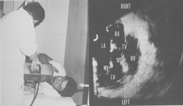
From No.3 Fig.5 Insertions of transducer to esophagus and transesophageal ultrasound examinations wer performed with patients in left lateral position(left). Typical horizontal scan in a 26-year-old normal adult by using transesophageal method (right).

From No.3 Fig.6 A long axis scan in a 31-year-old normal man. Entire heart image is seen. The endocardium of the left ventricle and the right ventricular anterior wall are seen.

From No.3 Fig .9 Transesophageal M-mode echograms. These images were recorded in order to identify echo sources of transesophageal cross-sectional images shown in Fig.10. Arrows A,B and C of Fig.10A and Fig.10D correspond to Fig.9A, 9B and 9C in each and show directions of M-mode echograms.

From No.4 Transesophageal horizontal scan at the level of aortic valve in a normal adult.

Photography of original paper

From No.12 Fig.2 Trans-stomach-wall high speed rotating scanner.

From No.12 Fig.3 Horizontal scan through the left kidney in a normal adult by using the trans-stomach-wall rotating scanner. When near gain is standard. Pancreas is seen as echo free space near the esophagus.

From No.12A Transgastric high speed rotating scanner with flexible tube.

From No.12A Horizontal scan at the level of kidney in a normal adult. Pancreas is seen very clearly.

From No.13 Transesophageal high speed rotating scanner.

From No.13 Transducer and commutator in oil bag.

From No.13 Horizontal scan at the level of the mitral valve in a patient with severe mitral stenosis by using the transesophageal high speed rotating scanner. Anterior and posterior mitral leaflets are thickened.

From No.14 Fig.1 Preoperative horizontal scan at the level of the mitral valve in a patient with atrial septal defect by using a transesophageal high speed sector scanner. ( case 1) D = defect.

From No.14 Fig.3 postoperative horizontal scan at the level of the mitral valve in the patient as show in Fig.1 and2 by using a transesophageal high speed rotating scanner (case 1).

Photography of original paper

From No.16 Fig.1 Diagrammatic illustration of the transgastric ultrasound sector scanner with gastrofiberscope.

From No.16 Fig.2 Transgastric ultrasound sector scanner with gastrofiberscope (Olympus GIF type Q).

From No.16 Fig.3 Horizontal scan through the stomach posterior wall in a normal adult by using the transgastric sector scanner with gastrofiberscope when the stomach was filled with water. Left kidney is seen clearly. SW = stomach wall, LK = left kidney, V = vertebra, R = right, L = left.

From No.17 Fig.5 Transesophageal inferior oblique scan through the apex in a normal adult. Cross-section is angled downward about 40 degrees from the horizontal plane. The entire heart including the apex is seen.

From No.18 Transesophageal horizontal sweeping in a normal adult at the level of mitral valve.
From No.18 Transesophageal horizontal sweeping in a normal adult at the level of aortic valve.

From No. 21 Upper Fig. Intragastric high speed rotating scanner. Small transducer in stomach is rotated by flexible rotating shaft and motor at 15-50 cycles/sec. Lower Fig. Transducer and commutator in oil bag.


Left Right
From No.21 Horizontal scans through left kidney in normal adult with intragastric high speed rotating scanner.
Left : Left kidney and abdominal aorta are seen clearly. If amplitude of near field is relatively low, pancreas is seen as anehoic
space near stomach wall. Right : With increasing amplitude of near field, pancreas assumes cloudlike shape.

From No.22 Fig. 4 Transesophageal cross-sectional echocardiogram before surgical repair in a patient with Lutembacher's
syndrome.. Cross-section is horizontal. ESO = esophagus, D = defect, LA = left atrium, LV = left ventricle, RA = right atrium, RV = right ventricle.

From No.22 Fig.5 Transesophageal cross-sectional echocardiogram after surgical repair in a patient with Lutembacher's syndrome. Cross-section is horizontal. Interatrial septum is seen continuously . IAS = interatrial septum.

From No.24 Transesphageal horizontal scan at the level of the aortic valve . The aortic cusps are closed in diastole. Large left atrium is seen . Right ventricular outflow tract is seen anterior to the aorta. AV = aortic valve, RVOT = right ventricular outflow tract.

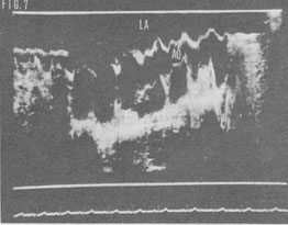
From No.24 Trans esophageal vertical linear scan through the pulmonary artery in a normal subject. Bifurcation of pulmonary artery and part of ascending aorta are seen. AO = aorta, PA = pulmonary artery , BI = bifurcation of pulmonary artery.

From No.25 Diagrammatic illustration of the transesophageal high speed rotating scanner. A small transducer in the esophagus is rotated through a full 360° through a flexible shaft by a motor at 15 to 50 cycles /s. Although the small trasducer is rotated with great speed in the patient's esophagus, no damage results because the transducer is safely enveloped in an oil bag.

From No.25 Transesophageal high speed rotating scanner.

From No.25 Transducer and commutator in oil bag. Sound energy is coupled to and from the transducer through the slip-ring commutator because of the full 360° rotation of the transducer.


From No.25 Transesophageal cross-sectional echocardiograms in a patient with mitral stenosis. The cross section is horizontal and shows the heart as viered from the cardiac apex. A : a frame during diastole and B : a frame during systole. A stenotic mitral orifice(in A) is seen between the tips of the thickened mitral leaflets. The interatrial septum (IAS) is seen without dropout. AML = anterior mitral leaflet. ESO = esoophagus, IVS = interventricular septum, LA = left atrium, LV = left ventricle, PML = posterior mitral leaflet, RA = right atrium, RV = right ventricle, TV = tricuspid valve.

From No. 26 Fig.2 Transesophageal two-dimensional echocardiogram in a patient with an ostium secundum atrial septal defect. Cross-section is horizontal. D - defect.

From No.27 Transesophageal two-dimensional echocardiogram in a patient with a left atrial myxoma. Cross section is horizontal. A stalk of the tumor is seen clearly. K = stalk.

From No.27 Fig.5 Extracted tumor. Weight of the tumor was 38.5g.

From No.28 Transesophageal two-dimensional echocardiogram in a patient with both aortic regurgitation and mitral stenosis. LCA = left coronary artery, RCA = right coronary artery.

From No.29 Fig.6 Horizontal scan in a normal adult. A part of aorta (A) is seen. There is this paper in internet. In internet, as references of my paper appear at first, please scroll down, then my full paper will appear. Please click here

From No.30 FIg.2 Traansesophageal two-dimensional echocardiogram of a patient with mitral stenosis. Image shown by arrow A is probably left coronary artery.